New crystal structures of translocating ribosome
Several new crystal structures of the 70S ribosome in complex with EFG and non-hydrolyzable GTP analogs have revealed how the ribosome directionally translocates mRNA and the tRNAs through the A, P, and E sites and how specific features of EFG and ribosomal RNA act as pawls to enforce this ratcheting mechanism. The new structures were solved in the Cate, Noller, and Ramakrishnan labs and were published in the June 28 edition of Science. Many of the new structures were made possible by data collected at the SIBYLS beamline.
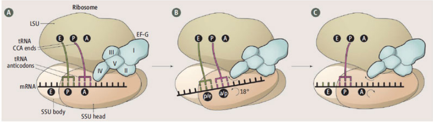
If you want the boiled down version of these new results then read this succinct comment by Marina Rodnina.
Rodnina MV. “Translocation in action.” Science. 2013 Jun 28;340(6140):1534-5.
A figure liberally lifted from her comment.

Tourigny DS, Fernandez IS, Kelley AC, Ramakrishnan V. “Elongation Factor G Bound to the Ribosome in an Intermediate State of Translocation.” Science. 2013 Jun 27;340(6140)
Pulk A, Cate JHD. “Control of ribosomal subunit rotation by elongation factor G.” Science. 2013 Jun 28;340(6140).
Zhou J, Lancaster L, Donohue JP, Noller HF. “Crystal structures of EF-G-ribosome complexes trapped in intermediate states of translocation.” Science. 2013 Jun 28;340(6140).